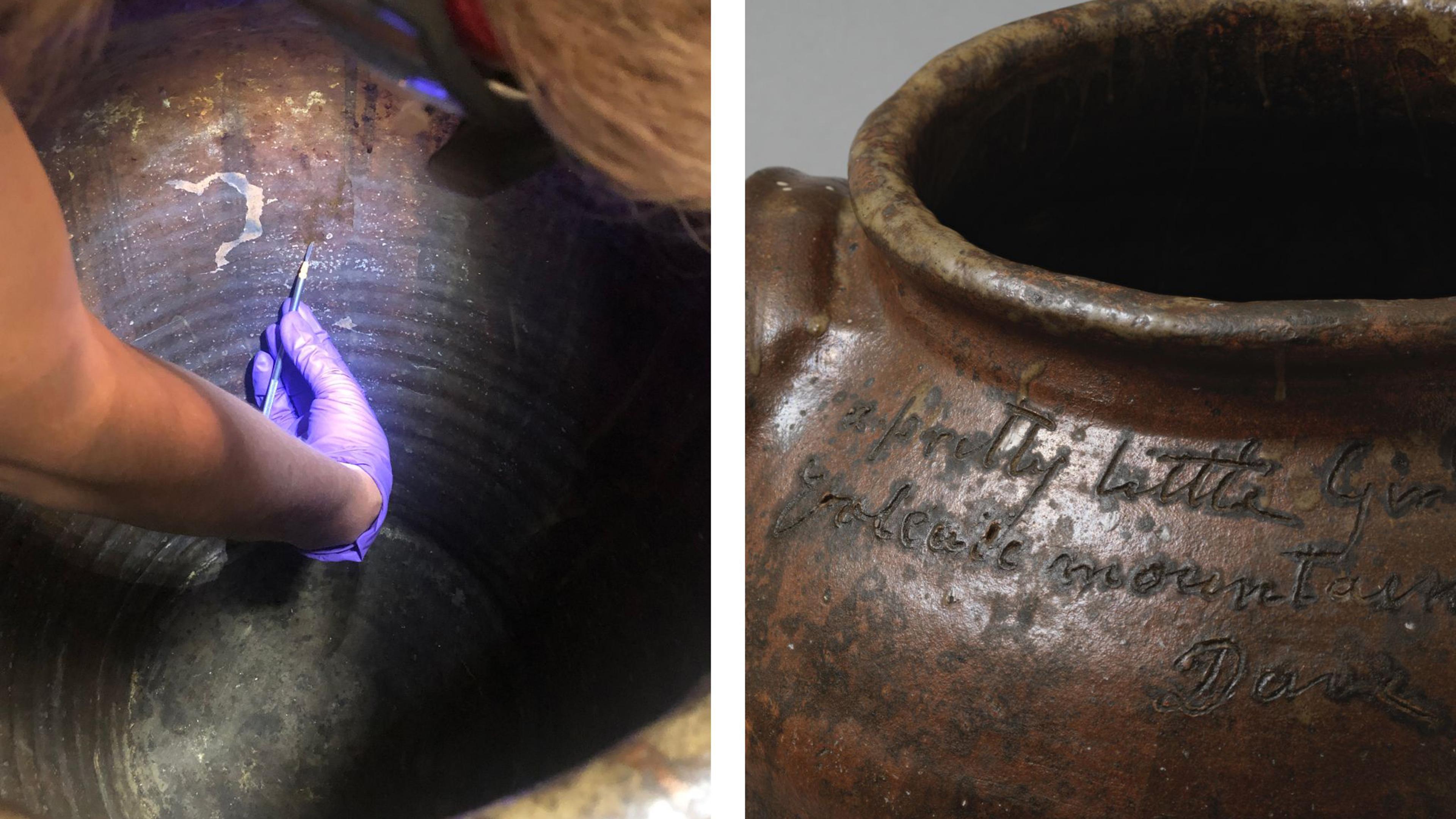
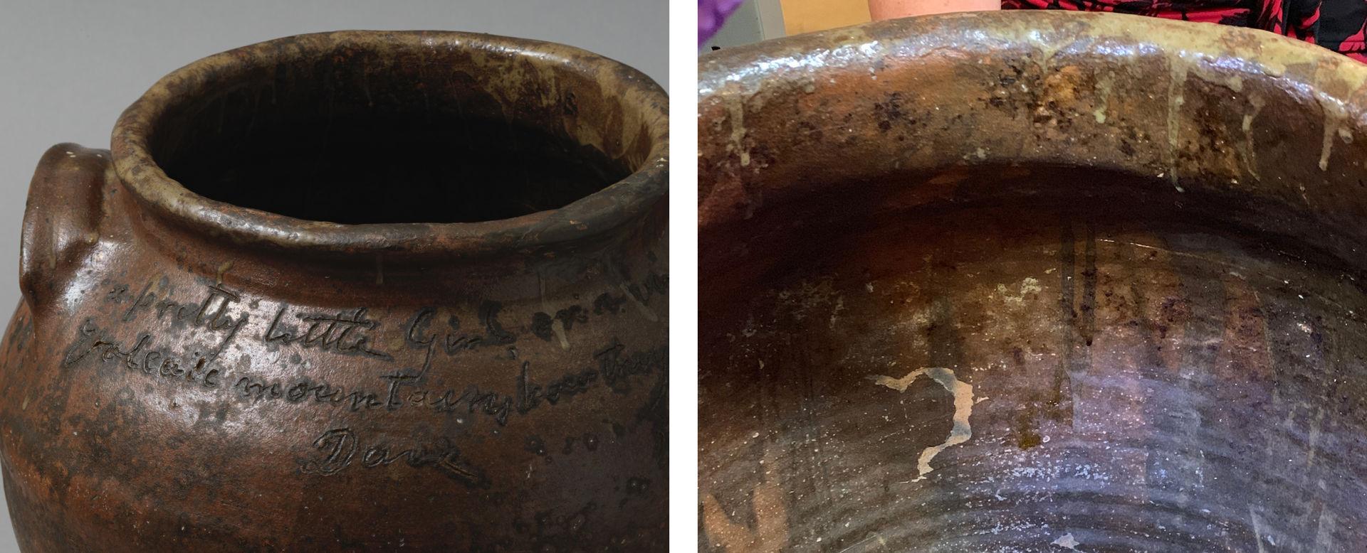
McKissick museum jar 5.100 with exterior drips and view inside of the vessel
The exceptional and exciting occurrence of organic food residues inside large nineteenth century alkaline-glazed stoneware vessels from the Old Edgefield district, some also signed by enslaved potter and poet David Drake, has offered a unique opportunity to study those residues as anthropological evidence of the jars’ contents (e.g., preserved meat, fat, eggs, etc.), potentially offering information on the use of the jars and the lifestyle of the people that used them. Residues scraped from the jars’ surfaces were analyzed in the Department of Scientific Research of The Met to evaluate their composition, assess contaminations and clues to their condition (e.g., presence and extent of degradation products), all informing of the next process to further characterize the residue. A selection of samples was also sent for analysis by our ARCHE collaborators at the University of Bordeaux, so that tissue or species sources could be identified, and if age or processing information was attainable.
When tackling analysis of historic food residues, analysts must grapple with variables potentially affecting the residues’ composition and integrity. Where on unglazed earthenware, organic residues can be preserved, absorbed into the porous ceramic fabric, residues on pottery glazed or fired at high temperature remain exposed on the surface and are more vulnerable to compositional changes due to washing and cleaning, as well as contamination.
Materials deposited from smokerooms, animals (e.g., insects and other critters)-, and human- interaction (skin cells, hair, clothing fibers), including those from reuse, contribute to environmental clues, but contaminate evidence of the jars’s historic content.
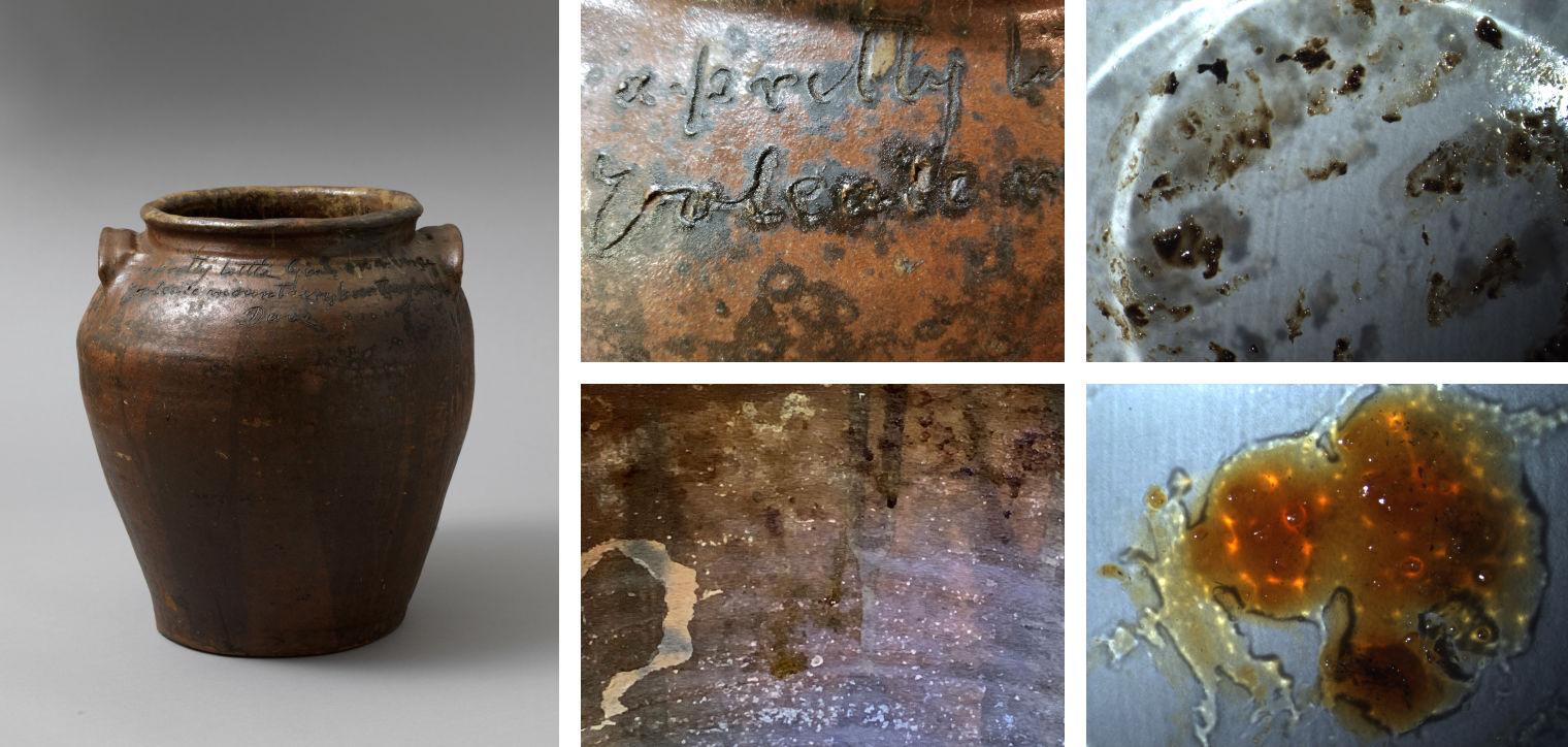
Figure 1. Left: Dave (later recorded as David Drake) (American, ca. 1801–1870s). Storage Jar (Inscription: verso: “a pretty little girl on a verge/ volca[n]ic mountains, how they burge”), 1857. Alkaline-glazed stoneware, Height: 19 in. (48.3 cm); diameter: 54 in. (137.2 cm); circumference, approximately 15-17 gallons. Donated by Belle Williams in memory of Admiral Yancy S. Williams. McKissick Museum, University of South Carolina (5.100). Top center and top right: detail of the location and appearance of the organic residues around the inscription. Bottom center and bottom right: detail of the location and appearance of the organic residues appearing as drips in the interior walls.
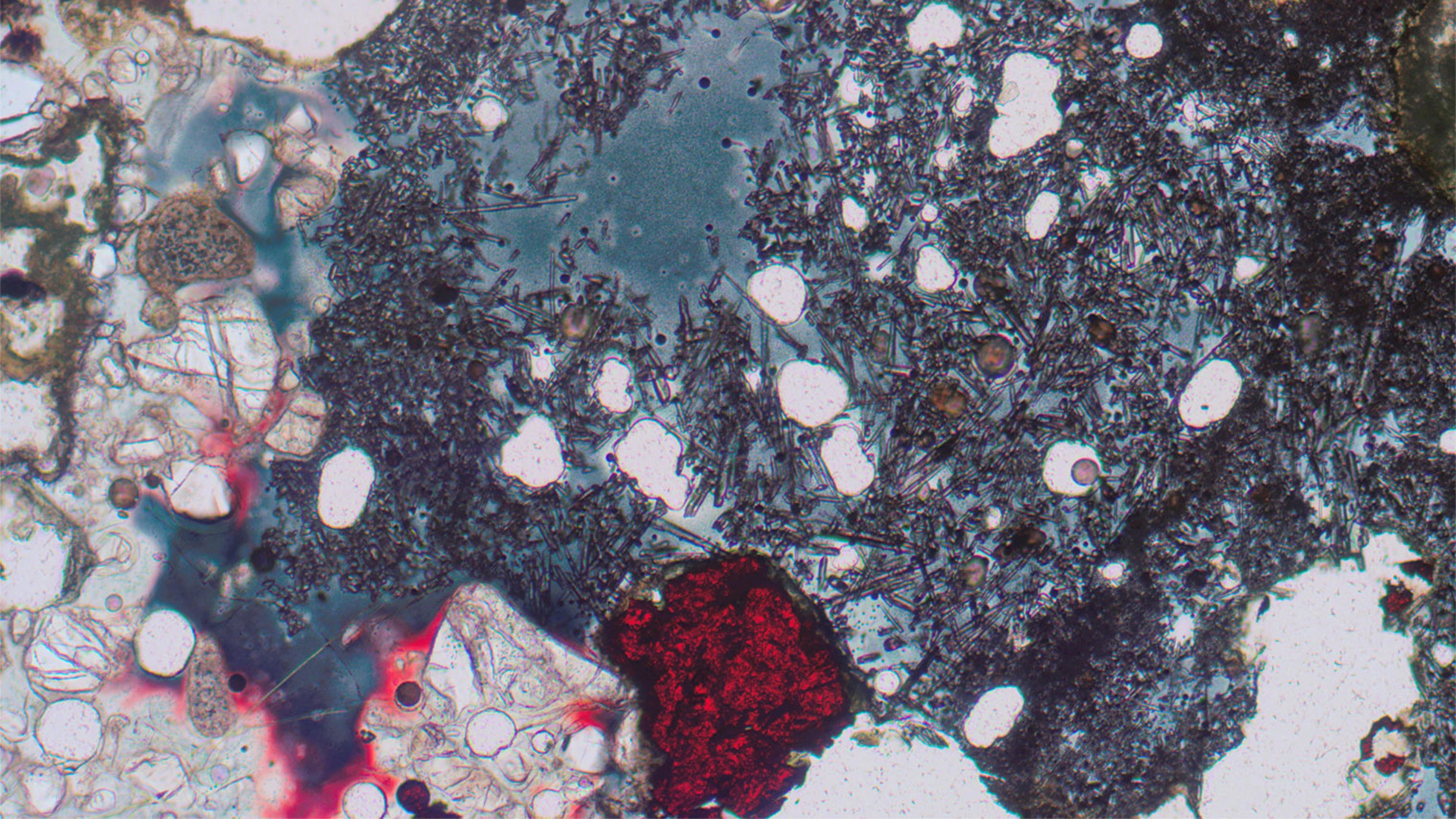
The samples varied in homogeneity and texture (viscous, jelly-like, gummy), color (clear, pale yellow, orange and black) and solids (quartz, black particles, fibers, animal hair, paint, insect parts). At the microscope, the solids which would have interfered with analysis were separated.
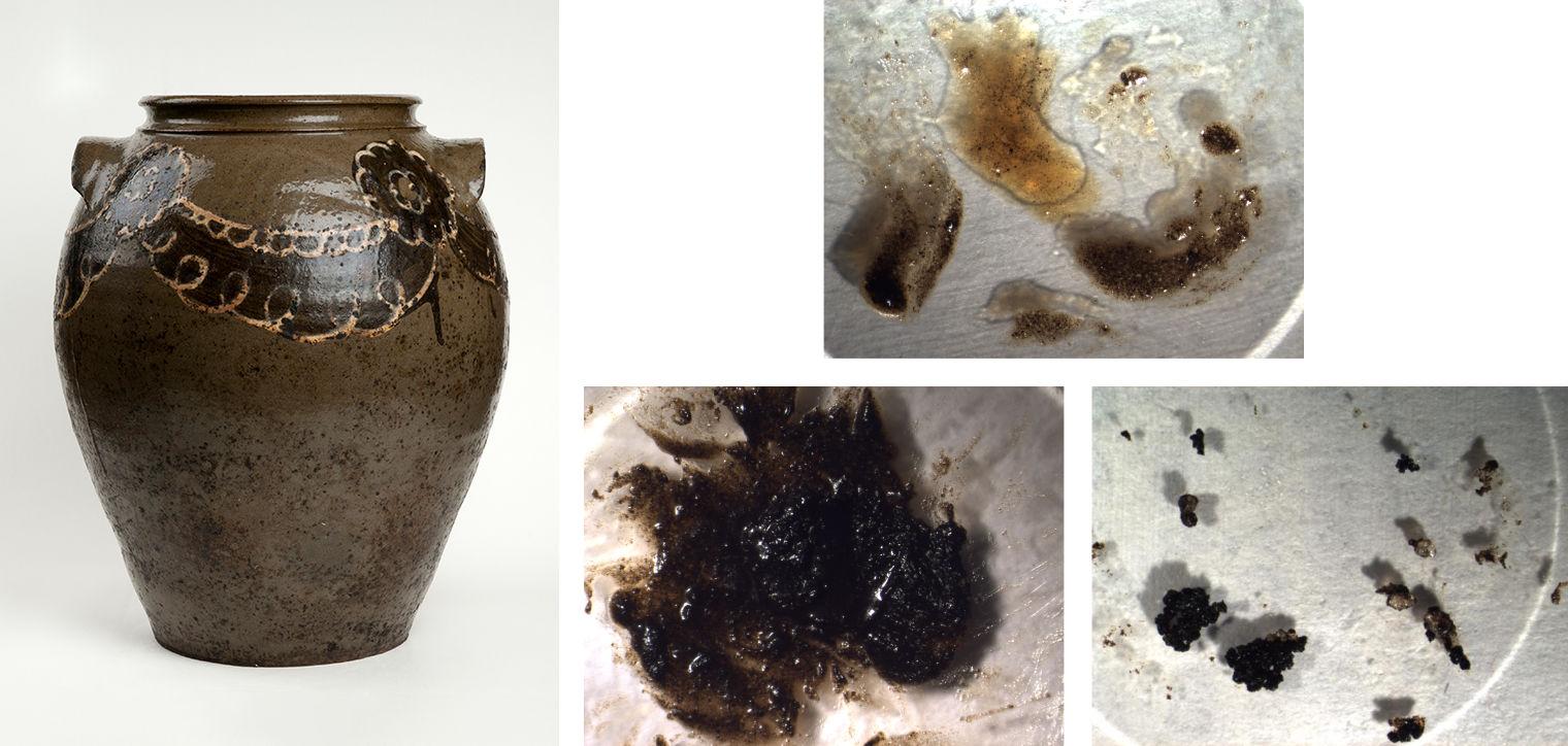
Figure 2. Left: Dave (later recorded as David Drake) (American, ca. 1801–1870s). Storage Jar, 1845. Alkaline-glazed stoneware, Height: 16 5/8 in. (42.2 cm); diameter: 7 5/8 in. (19.7 cm) ; circumference: 43 1/4 in. (109.8 cm). McKissick Museum, University of South Carolina (5.99). Top center: Photo-micrographs of the viscous fraction obtained after mechanical separation of the solid and jelly inclusions. Bottom center and right: jelly fraction with small carbon –based black inclusions, and larger black particles with quartz inclusions separated from the bulk residue, respectively.
Fourier transform infrared micro-spectroscopy (micro-FTIR), revealed that the samples are predominantly of lipidic (fat/oil) composition, confirming results obtained in situ with the portable FTIR at the McKissick Museum, Columbia, SC.
Within the miniscule samples used for this micro-analysis, only very few contained protein, in addition to lipids: gummy residues, such as those sampled from the lower areas of the jars’ interiors and under the handle of a jar. Most likely protected from the regular washing of the jar’s interior, the residues under the handle are also most contaminated from moving the jars with bare hands.
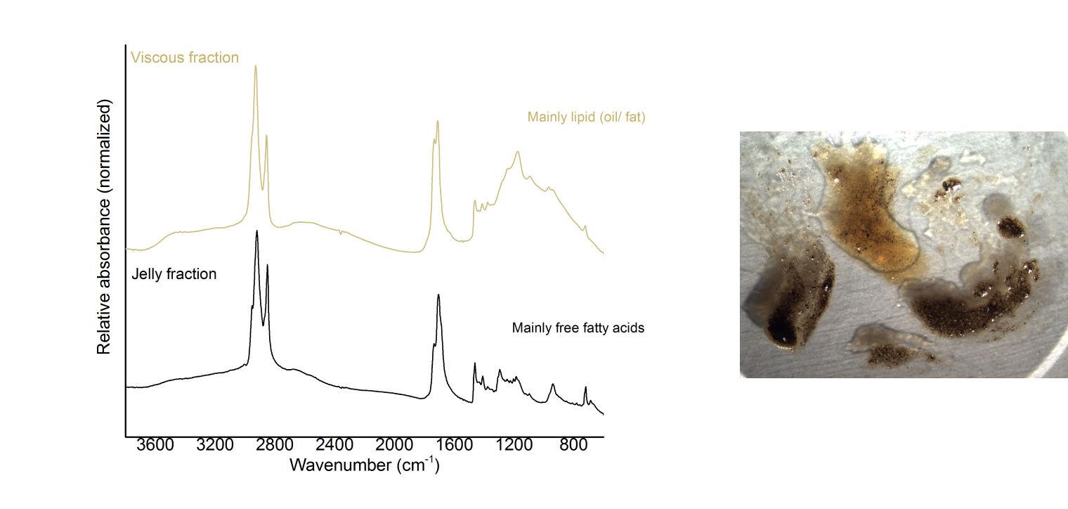
Figure 3. Comparison of FTIR spectra obtained from the two fractions (viscous and jelly) of the interior residue. The viscous fraction shows a characteristic profile for lipids, while the jelly fraction shows a characteristic profile for free fatty acids (mainly palmitic and stearic acids).
Lipids from fats or meat, compared to water-soluble proteins, could better survive the jars’ periodical washings in scalding water. However, they are altered by moisture and air, through pathways of hydrolysis and oxidation, with some of these processes having already taken place in the processing of lipids (e.g., fat rendering) and preservation of meat (brining, dry-curing).
Free fatty acids from hydrolysis of triglycerides, such as palmitic and stearic acid (both very abundant in animal fats), were indeed detected in the clear jelly-like and crystalline clusters inclusions, but also in the gummy black accumulations under a jar’s handle, and bottom of the jars.
Brownish inclusions in some of the residues were rich in metal oxalates. Oxalic acid, a degradation product of air-exposed organic matter (including proteins and lipids) and a metabolite of fungal activity, had formed salts with calcium, iron and other reactive ions likely originating from the residue, environmental particulate, and/or the jar’s exposed fabric and glaze components.
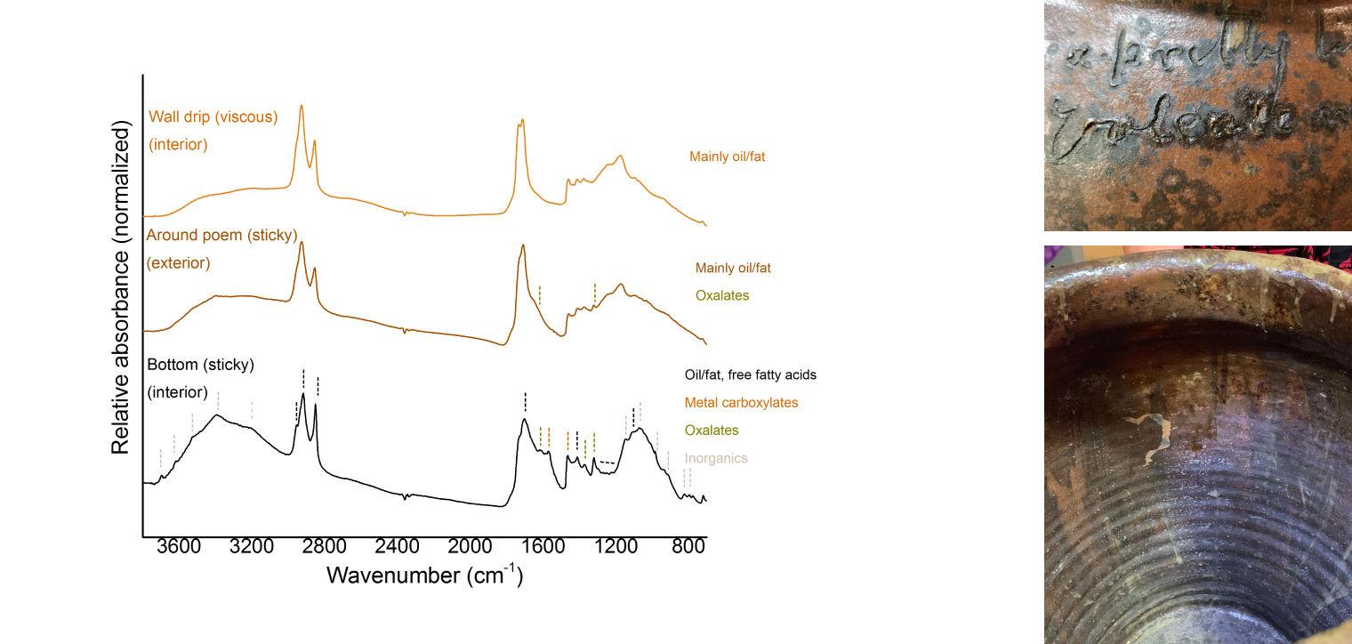
Figure 4. Comparison of FTIR spectra obtained from different locations of Dave’s jar (5.100) showing variability in compositions.
The extent of lipid hydrolysis in the residues was assessed by Evolved Gas Analysis-Mass Spectrometry (EGA-MS). By this technique, the residues were heated in a miniature furnace over a temperature gradient (100 to 700oC) and their constituents detected by the mass spectrometer in function of the temperature and time ranges at which they evolved from the sample. The temperature profiles (thermograms) obtained show the distribution of the volatile fractions (e.g. free fatty acids) and less volatile (triglycerides/ diglycerides) fraction within the sample. Their relative ratios give a sense of the hydrolysis undergone by the residue.
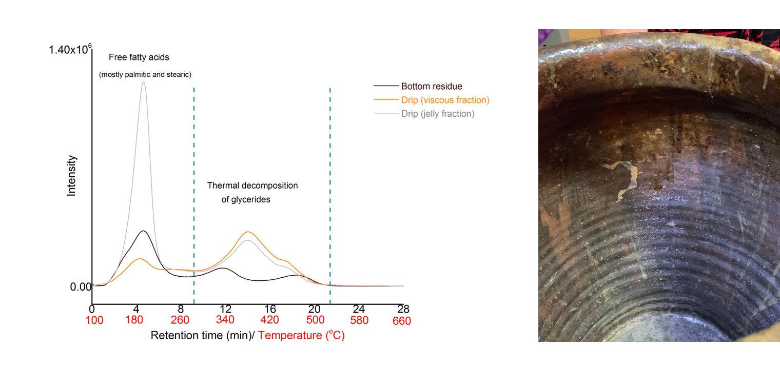
Figure 5. Comparison of thermograms obtained from evolved gas analysis-mass spectrometry (EGA-MS) of residue from the wall drip (orange and gray trace) and from the bottom (brown trace), all from the interior of Dave’s jar (5.100), showing variability in compositions, with material losses for free fatty acids up to 280⁰C, followed by decomposition of glycerides from ca. 300⁰C. The jelly fraction from the wall drip is more rich in free fatty acids, mainly palmitic and stearic acids, highlighting the accumulation of free fatty acids from lipid hydrolysis into jelly inclusions.
The identity of the individual fatty acids, free or bound in glycerides, as well as the products of oxidation of the unsaturated fatty acids, was assessed by Thermally assisted Hydrolysis and Methylation Gas – Chromatography-Mass Spectrometry (THM-GCMS). Here the lipid residue is hydrolyzed and hydroxyl groups (-OH) from fatty acids and glycerol are methylated and identified by GCMS, providing distributions of fatty acids, some of which attributable to animal sources. In addition, oxidized fatty acids derived from unsaturated fatty acids, as well as dicarboxylic acids, although not specific to origin of the lipids, are helpful clues to the oxidation of the residue analyzed.
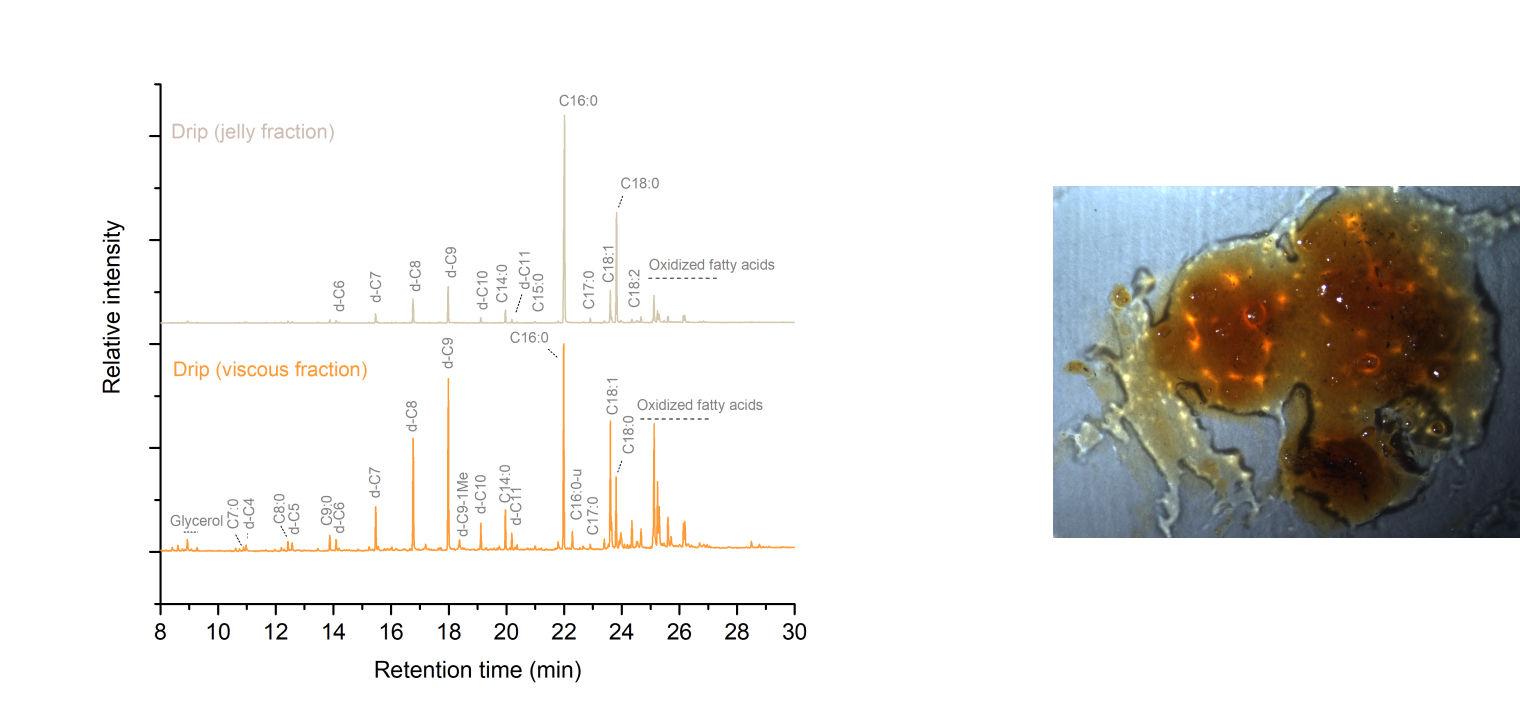
Figure 6. Comparison of chromatograms obtained from THM-GCMS analysis of residue from the jelly and viscous fractions of the wall drip inside Dave’s jar (5.100), showing variability in distribution of fatty acids, with relatively higher amounts of palmitic and stearic acids (C16:0 and C18:0, respectively) in the jelly fraction, as well as relatively high amounts of oxidized fatty acids in both fractions. Saturated fatty acids methylesters: C7:0-C18:0, of which C15:0 and C17:0, indicative of animal fats. Dicarboxylic fatty acids di-methylesters: d-C4- d-C11, where d-C8 and d-C9 are suberic and azelaic acids, respectively, as oxidation products of fat/oil. Unsaturated fatty acids: C18:1 and C18:2 are mono and di-unsaturated C18:0, where C18:1 is present in high amounts in fats. Partially methylated fatty acid: d-C9-1Me. Underivatized: C16:0-u.
With minimal sample sizes required for the analyses described, the organic composition of the residues can be interpreted as likely including animal and possibly plant lipids with occasional protein, as well as degradation products, but there still leaves unanswered the question: what exactly was stored in these jars? To begin to unravel the question of animal species source or food processing, we have to turn to more sophisticated “omics” techniques with our ARCHE collaborations at the University of Bordeaux, France.
The organic residues are an invaluable source for insight into these jars’s content and use, with their analysis and interpretation challenged by the chemical effects of curing, rendering, and preservation methods of meats or fats followed by aging during storage, and after the jars became collectable objects. Fat-rendering and meat preservation processes were replicated in the lab in small scale to eventually address these questions, but there remain the challenges, both analytical and logistical, to pursue such investigation in a museum lab and underscores the necessity for collaborative efforts. Nevertheless, it is hoped that broader discussions about the importance of this research focused on Old Edgefield’s jars stimulate further studies on these and other historical residues, to the benefit of the field of science, anthropology and food history.