A Survey of the Microcosme or the Anatomie of the Bodies of Man and Woman wherein the Skin, Veins, Nerves, Muscles, Bones, Sinews and Ligaments Thereof are Accurately Delineated, and so Disposed by Pasting, as that Each Part of the Said Bodies Both Inward and Outward are Exactly Represented. Useful for all Doctors, Chyrurgeons, Statuaries, Painters, &c.
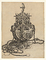
The anatomical image with attached moveable flaps that, when lifted, reveal underlying layers that illustrate the organs, blood vessels, and bones of the human body is a type of print that was produced throughout the sixteenth and seventeeth century. The many different language editions and reeditions testify to the popularity of this genre. The tradition goes back to Strasbourg artist Heinrich Vogtherr’s depiction of a seated woman of 1538 in which a flap on the woman’s belly can be lifted to reveal the organs inside.
The present piece illustrates the continuation of that tradition into the seventeenth century in England. The sheets originally formed a book that was later disbound. The work includes the title page, four engravings, and four pages of letterpress text explanations of the diagrams. The work was published by Joseph Moxon, a British publisher and mathematician, and dedicated to Samuel Pepys (1633-1703), best known for his diary, an important primary source about the period.
The present piece illustrates the continuation of that tradition into the seventeenth century in England. The sheets originally formed a book that was later disbound. The work includes the title page, four engravings, and four pages of letterpress text explanations of the diagrams. The work was published by Joseph Moxon, a British publisher and mathematician, and dedicated to Samuel Pepys (1633-1703), best known for his diary, an important primary source about the period.
Artwork Details
- Title: A Survey of the Microcosme or the Anatomie of the Bodies of Man and Woman wherein the Skin, Veins, Nerves, Muscles, Bones, Sinews and Ligaments Thereof are Accurately Delineated, and so Disposed by Pasting, as that Each Part of the Said Bodies Both Inward and Outward are Exactly Represented. Useful for all Doctors, Chyrurgeons, Statuaries, Painters, &c.
- Artist: Johann Remmelin (German, 1583–1632)
- Artist: Michael Spaher (German)
- Publisher: Joseph Moxon (British, 1627–1691)
- Translator: John Ireton
- Dedicatee: Samuel Pepys (British, 1633–1703)
- Date: 1675
- Medium: Engraving and letterpress text
- Dimensions: Sheet: 15 1/4 × 12 1/4 in. (38.7 × 31.1 cm)
- Classification: Prints
- Credit Line: Mary Oenslager Fund, 2018
- Object Number: 2018.266a–f
- Curatorial Department: Drawings and Prints
More Artwork
A slider containing 5 items.
Press the down key to skip to the last item.
Press the down key to skip to the last item.

German
1750–70
Martin Schongauer
After/with Georg Schongauer
1470–1491
German
ca. 1590

Albrecht Dürer
1514
German
ca. 1620–23
Research Resources
The Met provides unparalleled resources for research and welcomes an international community of students and scholars. The Met's Open Access API is where creators and researchers can connect to the The Met collection. Open Access data and public domain images are available for unrestricted commercial and noncommercial use without permission or fee.
To request images under copyright and other restrictions, please use this Image Request form.
Feedback
We continue to research and examine historical and cultural context for objects in The Met collection. If you have comments or questions about this object record, please contact us using the form below. The Museum looks forward to receiving your comments.